


.")
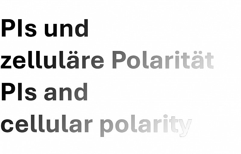
Cell polarity, or the establishment of asymmetries along an axis of polarity, is the basis for the development of complex multicellular tissues in all higher organisms. One approach to study the regulation of cell polarity is to look at specialized cell types that display extreme forms of polar tip-growth. Examples for plant cells with such properties are pollen tubes, root hairs or trichomes, which all represent single cells. The analysis of Arabidopsis mutants with various defects in PI metabolism revealed that PIs control vesicle trafficking and the actin cytoskeleton, which are aspects relevant for the manifestation of polarity. Current work is aimed at understanding how PIs control polar tip-growth. A particular focus area is the question how different regulatory effects of PIs on the cytoskeleton, vesicle trafficking and secretion are coordinated within a tip-growing cell.
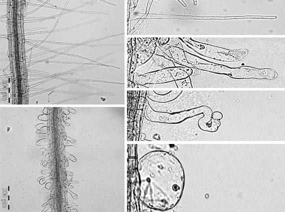
Effects of under- or overexpressing a PI4P 5-kinase on development and polarity of root hairs in Arabidopsis thaliana. Left, Wild type (top) and pip5k3-4 mutant (bottom). Right, Wild type (top) and progressive loss of polarity in root hairs with increasing levels of expression. (from Stenzel et al., Plant Cell 20, 124-141, 2008)
Cell polarity can manifest not only in the shape of a cell but also in the asymmetric distribution of subcellular components. The polar distribution of various proteins is a prerequisite the function of cells in the context of a tissue. A prominent example are the auxin transporters of the PIN-FORMED-family (PIN proteins), which mediate the export of the phytohormone, auxin, across the plasma membrane. Depending on to which side PIN proteins localize in adjacent cells, auxin is distributed directionally within a tissue. For instance, in the Arabidopsis root tip the interplay of various mainly basally or apically localized PIN proteins results in the formation of an auxin gradient, which serves as a positional cue for differentiation of cells and the development of different tissues. PIs serve as additional positional signals, guiding the distribution of PIN proteins. Current work is aimed at elucidating how PIs mechanistically contribute to the control of PIN distribution. A particular emphasis is given to the understanding how PIs themselves are distributed in a polar fashion.
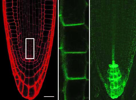
Polar distribution of PIN1:GFP in root cells of Arabidopsis thaliana. Left, Root tip stained with propidium iodide; center, PIN1:GFP-fluorescence in cells of the central cylinder (also see white box, left); right, auxin gradient visualized using a DR5:GFP-reporter. (modified from Ischebeck et al., Plant Cell 25, 4894-4911, 2013)